지난 연제들을 통해 GBR 및 Growth Factors에 대해 알아봤다. 이번 연제와 다음 연제를 통해 2017년 11월 9일에서 11일간 미국 시카고에서 열린 2017 World Workshop 의 내용 중, Work group 4에서 다룬 ‘PERI-IMPLANT DISEASES AND CONDITIONS’에 대해 살펴보고자 한다.
GBR 및 Growth Factors를 통해 Alveolar Bone을 잘 만들어 놨다고 해도, 추후 Implant 의 식립 및 Long-term Predictability가 중요한 문제이기 때문에 이에 대해 명확하게 알고자 최신 지견을 찾아보던 중, 미국의 American Academy of Periodontology(AAP)와 유럽의 The European Federation of Periodontology(EFP)가 후원하고, Journal of Clinical Periodontology(JCP)와 Journal of Periodontology(JOP)에 2018년 6월에 동시 게재됐으며 전 세계의 유명한 연자 및 교수님들이 모여 Workshop을 해 만든 2017 WWP (2017 World Workshop proceedings)에 대해 알아보는 것이 큰 의미가 있다고 판단했다.
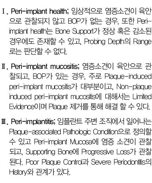
Work group 4에서는 ‘PERI-IMPLANT DISEASES AND CONDITIONS’에 대해 다뤘는데, Periimplant Health, Periimplant Mucositis 그리고 Periimplantitis라는 새로운 분류를 도입했고, 아래와 같이 정리했다.
첫 번째로, Peri-implant Health에 대해 논의한 내용들을 알아보자.
Microscopic Level로 보면, Healthy Peri-implant Mucosa는 안쪽에 Connective Tissue를 Keratinized Epithelium(masticatory mucosa)나 Non-keratinized Mucosa(Lining mucosa)로 덮고 있고, 3~4mm 정도의 Height를 가지는데, 약 2mm의 Epithelium을 가지고 Inflammatory Cells의 Cluster가 Connective Tissue에 분포한다. 임플란트의 Intrabony Part는 60% 정도는 Mineralized Bone과 접촉해 있고, 나머지 부위는 Bone Marrow 쪽에 있는 것으로 관찰된다. 임플란트와 접촉하는 Peri-implant Mucosa는 Coronal 쪽의 Junctional Epithelium과 Apical 쪽의 Sulcular Epithelium으로 구성되는데 Sulcular Epithelium 쪽에 주로 Vascular Plexus 등이 분포하고 있고, Masticatory Mucosa 쪽에 Crestal Bone에서 임플란트 표면에 평행하게 주행하는 Collagen들이 Main으로 분포하며 Circular Fiber들도 존재하는 것으로 보인다. Electron scanning microscope 상에서 CT의 200um Zone을 관찰해 보면, Inner Layer 40um에는 많은 Fibroblast들이 관찰되고, Outer Layer 160um에는 Collagen Fiber(83%)와 약간의 Fibroblast 및 혈관들이 관찰됐다.
사실, 필자가 가장 궁금해 했던 Mucosal Adhesion의 Morphogenesis에 대해서는 간단히 다뤘는데, 3개월간의 Healing을 Biopsy를 통해 관찰한 결과, 초기 힐링단계에서 CT에 Neutrophils와 Macrophages로 구성된 Fibrin Clot/coagulum이 형성되고 2~3주 지나면 Fibroblast 수는 감소하고 Collagen Matrix가 증가하기 시작하며 oral epi.로부터 이동한 Epithelial Cells가 CT의 Marginal Part를 차지하기 시작한다.
6~8주 지나면 Mucosal Adhesion이 Maturation되고 Tissue-implant의 Interface Zone은 Epithelial과 Connective Tissue Adhesion의 Combination으로 구성된다.
Peri-implant Mucosa의 Dimension 즉, Biological Width는 동물실험들에서 관찰한 결과로는 2mm 정도의 Epithelium과 1~2mm 정도의 Connective Tissue로 구성된다고 볼 수 있고, Mini-implant를 이용한 Human Studies에서는 치유될수록 Mucosa의 Height가 길어지는데, 2주차때 2.7mm 정도에서 4~12주차가 되면 3.5mm 정도까지 되고, Epithelium이 2~2.2mm, CT가 1.1~1.7mm 정도 되는 것으로 관찰됐다. 즉, Peri-implant Mucosa의 Height는 3~4mm 정도이고, Epithelium이 2mm 약간 이상 정도라고 보면 될 것이다.
Bone Sounding이나 Transmucosal Sounding을 한 결과는 자연치나 임플란트 모두 Proximal에서 Facial/Buccal보다 더 깊게 나타났고, 자연치에서보다 임플란트에서 좀 더 깊은 것으로 관찰됐다. 이는 Root Cementum의 부재 및 Collagen Fiber의 방향이 원인으로 보인다.
Keratinized Mucosa는 Masticatory Mucosa로 Peri-implant Mucosa의 Margin에서 Movable Lining Mucosa까지를 말하는 것으로, 이 KM은 Lamina Propria와 Orthokeratinized Squamous Epithelium으로 덮여있는 조직으로 발치가 KM의 감소에 주 원인으로 보인다.
임플란트와 자연치의 주된 차이를 살펴보면, 임플란트에는 Root Cementum, PDL 그리고 Bundle Bone(alv. bone proper)의 부재로 Dento-alveolar, Dento-gingival fiber가 없으며 자연치에는 CEJ가 Gingiva의 Outline을 형성한다면, 임플란트에서는 Crestal Bone의 Contour 및 인접치의 CT 부착이 Gingival Contour에 영향을 미친다고 볼 수 있다.
두 번째, Peri-implant Mucositis에 대한 논의에서는 Loss of Supporting Bone이나 Continuing Marginal Bone Loss가 없는 상황에서 Soft Tissue의 염증상황으로 정의했으며 ‘A Disruption of the Hostmicrobe Homeostasis at the Implantmucosa Interface and is a Reversible Condition at the Host Biomarker Level’이라고 표현한다.
즉, 가역적 상황이고, Biofilm Control을 통해 완전한 해결할 수 있다고 보고 있다. Peri-implant mucositis는 Connective Tissue 내의 Inflammatory Cell Infiltrate로 설명할 수 있으며, 3주 이상의 오래된 Peri-implant Mucositis 상황에서는 Inflammatory Cell Infiltrate의 영역이 커져있음을 관찰할 수 있었다. 주기적인 Supportive Peri-implant Therapy가 Peri-implantitis로 진행되는 것을 막을 수 있는 중요한 Preventive Treatment라고 설명했다.
여러 연구에 의하면 임플란트 식립 후 Healthy Peri-implant Mucosa 형성 후에, 3주간의 Oral Hygiene Practice를 중단하면 염증소견이 Peri-implant Mucosa에 발견되고 B cell과 T cell의 비율이 증가하며, Inflammatory Cell Infiltrate의 Size와 면역세포 수가 증가하는 것을 관찰할 수 있다. 어떤 연구에서는 3주간의 Oral Hygiene Practice를 자연치(Experimental Gingivitis)와 임플란트(Peri-implant Mucositis)에서 중단 시, 임플란트 부위에 Biofilm Accumulation이 자연치보다 덜하지만, 염증소견은 자연치에 비해 Peri-implant Mucosa 부위가 더 심한 것으로 밝혀졌다.
다음 연제를 통해 Periimplantitis, The Etiology of Hard and Softtissue Deficiencies at Dental Implants, Case Definitions and Diagnostic Considerations, Consensus Report 등에 대해 논의된 내용을 바탕으로 2017 WWP(2017 World Workshop proceedings)에 대해 알아보도록 하겠다.